Getting to the heart of the matter: 10 years of cardiovascular research funded by BSAVA PetSavers
9 May 2024
Being 50 years old is a time for BSAVA PetSavers to look back and consider its impact. Much has been achieved by the last 10 years of cardiovascular clinical research funding. Here, we focus in on two major cardiac diseases: hypertrophic cardiomyopathy (HCM) in cats and myxomatous mitral valve disease (MMVD) in dogs.
Hypertrophic cardiomyopathy (HCM)
Research suggests that over 1.5 million cats are affected with hypertrophic cardiomyopathy (HCM) in the UK alone. PetSavers’ funding has been part of larger funding which has transformed its diagnosis, making this easier for first opinion practitioners and leading to earlier treatment with potentially less distress.
HCM often affects relatively young cats, and can result in heart failure, respiratory distress, very painful arterial thromboembolisms and death. However, many cats with HCM remain asymptomatic and are at risk of being undetected until the rapid development of unexpected spontaneous or stress-induced signs. Veterinary visits and procedures can be the stress-inducing events causing these signs.
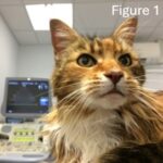
 Historically, asymptomatic cats with heart murmurs were diagnosed by referral cardiologists, but financial pressures led to many high-risk cats being undetected and unscreened. Professor Virginia Luis Fuentes and her RVC research team, aided by IDEXX, changed veterinary practice by developing first opinion, in-house screening techniques for HCM including blood NT-proBNP measurements and focussed echocardiology screens measuring the left atrial diameter.1,2 This ultrasound technique is now taught to vet students and primary care vets to be used as standard in first opinion veterinary practice (Figure 1). NT-proBNP screening tests are now also used widely in first opinion practice.
Vicky Ironside and colleagues carried out a PetSavers-funded study that looked at prognostic indicators for cats with asymptomatic HCM. They confirmed that left atrial enlargement and elevated N-proBNP both indicated an increased risk of a cardiac event, including congestive heart failure, arterial thromboembolism or sudden death. Increased speed of atrial enlargement elevated this risk.3 This study was awarded the BSAVA PetSavers Veterinary Achievement Award in 2021.
Cats showing respiratory distress and dyspnoea are critically ill, medically unstable and vulnerable to the effects of stress. Being held for blood sampling is an invasive, potentially stressful event for any cat. PetSavers funded two studies4,5 which demonstrated that N-proBNP concentrations in pleural fluid from dyspnoeic cats, being removed for therapeutic and diagnostic reasons, could be used to distinguish between cardiac and non-cardiac causes of dyspnoea in primary practice. This removes the need for blood sampling and aids rapid diagnosis and treatment without referral.
Dr Dave Dickson received joint PetSavers and Veterinary Cardiovascular Society (VCS) funding to confirm his initial research which developed the RAPID assessment for dyspnoeic cats.6 His earlier work showed that dyspnoeic cats presenting with hypothermia, tachycardia, gallop sounds or profound tachypnoea are likely to have dyspnoea from an underlying cardiac cause, although a confirmatory diagnosis needs to be made. We await the results of his further investigations.
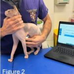
 Our support of research into HCM continues with other on-going studies under Dr Melanie Hezzell at the University of Bristol looking at the role of myocardial fibrosis in feline heart and kidney disease. Additionally, we recently awarded joint funding with the VCS to a project developing an intelligent stethoscope for detecting HCM (Figure 2). This will use an inbuilt AI algorithm to differentiate between heart murmurs caused by HCM and non-harmful, physiological murmurs.
“PetSavers gives the profession access to research conducted in general practice, something which is rare. We need to improve the knowledge and management of diseases seen in general practice…” Dr Dave Dickson, RCVS Recognised Specialist in Cardiology
Myxomatous mitral valve disease (MMVD)
MMVD is a chronic, degenerative disease which can lead to fatigue, life-long medication and veterinary support, a reduced quality of life and, in some cases, heart failure, respiratory distress and reduced life expectancy. It accounts for over 70% of canine cardiovascular disease, and is seen in about 4% of dogs at primary care practices in the UK. Of these, 30% go on to develop cardiovascular compromise and heart failure.
In the last 10 years, PetSavers has funded student studies which have helped elucidate the pathophysiology of MMVD, potentially enabling future diagnostics and treatment. These have also introduced undergraduates to clinical research and launched researcher careers through master’s qualifications. We have a number of exciting, larger on-going studies looking at treatments used in primary practice, and the genetics of MMVD.
Examples of smaller PetSavers-funded studies include an undergraduate project at the RVC showing that dogs with MMVD may develop reductions in packed cell volume as their disease progresses, though the differences described were relatively small.7
MMVD’s progression is associated with progressive endothelial vascular dysfunction. In his master’s degree funded by PetSavers at the University of Edinburgh, Dr Marco Mazzarella showed that isometric myography, a technique not previously used in dogs, is feasible to be used on arteries from dogs with MMVD to identify loss of endothelial-dependent relaxation.8 Marco has now moved onto a PhD researching the endothelial glycocalyx in MMVD.
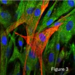
 Another student project funded by PetSavers in 2022, also at the University of Edinburgh, showed that senolytic drugs could reverse senescence while enhancing apoptosis in canine MMVD cultured valve interstitial cells (Figure 3), so might have potential as an extra therapeutic in MMVD treatment.
We have a number of larger studies in MMVD underway with Dr Hezzell’s research team. One is looking at how circulating fibrosis markers change with MMVD progression to help give additional insight into myocardial remodelling and potential design interventions to slow this. The second is investigating genes and pathways in MMVD in cavalier King Charles spaniels using a whole genome sequencing approach.
In 2016, the EPIC study demonstrated that small breed dogs with preclinical MMVD, managed with pimobendan in a specialist setting, remained in that preclinical stage for 15 months longer than small breed dogs given a placebo.9 In 2019, PetSavers funded Dr Elizabeth Bode and colleagues to investigate the impact of this study’s publication on pimobendan use in primary practice. Having demonstrated a marked increase in pimodendan prescription since 2016, they also found that most dogs prescribed pimobendan had not been assessed for cardiomegaly despite the fact that the EPIC study findings are only relevant to dogs with major cardiac remodelling. This suggests that some patients are likely to be having unnecessary prescriptions of this expensive drug.
“This was an ambitious project that combines the knowledge and specialisms of a number of clinicians and scientists, and which would not have been possible without the support of PetSavers and the Veterinary Cardiovascular Society.” Dr Elizabeth Bode, RCVS Specialist in Cardiology
At BSAVA PetSavers we are immensely proud that our funding has helped advance the care of pets with these major cardiovascular diseases. We have spent over £175,000 on clinical research into cardiovascular disease alone in the last 10 years. To fund ground-breaking research like that done by the researchers mentioned above, we rely on the generosity of our donors, supporters, fundraisers, legators and sponsors.
Please support us to continue this life changing work for pets by visiting the donation page on our website: https://www.bsava.com/petsavers/donate/
Figure 1. Cat about to be scanned using an echo machine.
Figure 2. Detecting HCM using a stethoscope.
Figure 3. Primary culture of mitral valves from dogs stained for markers of cellular senescence and apoptosis following treatment with senolytic drugs.
References
Sargent J, Connolly D, Watts V, et al. (2015) Assessment of mitral regurgitation in dogs: comparison of echocardiography with magnetic resonance imaging. J Small Anim Pract 56(11):641-50
Borgeat K, Dudhia J, Luis Fuentes V, et al. (2015) Circulating concentrations of a marker of type I collagen metabolism are associated with hypertrophic cardiomyopathy mutation status in ragdoll cats. J Small Anim Pract 56(6):360-5
Ironside V, Tricklebank P and Boswood A (2021) Risk indicators in cats with preclinical hypertrophic cardiomyopathy: a prospective cohort study. J Feline Med Surg 23(2):149-59
Hezzell M, Rush J, Humm K, et al. (2016) Differentiation of cardiac from non-cardiac pleural effusions in cats using second generation quantitative and point-of-care NT-proBNP measurements. J Vet Intern Med 30(2):536-42
Humm K, Hezzell M, Sargent J, et al. (2013) Differentiating between feline pleural effusions of cardiac and non-cardiac origin using pleural fluid NT-proBNP concentrations. J Small Anim Pract 54(12):656-61
Dickson D, Little C, Harris J, et al. (2017) Rapid assessment with physical examination in dyspnoeic cats: the RAPID Cat Study. J Small Anim Pract 59(2):75-84
Wilshaw J, Stein M, Lotter N, et al. (2021) The effect of myxomatous mitral valve disease severity on packed cell volume in dogs. J Small Anim Pract 62(6):428-36
Mazzarella M, Jones N and Culshaw G (2023) Isometric myography is a feasible method to identify and quantify endothelial dysfunction in dogs with myxomatous mitral valve disease. Am J Vet Res Sep 84(12):1-7
Boswood A, Haggstrom J, Gordon SG, et al. (2016) Effect of pimobendan in dogs with preclinical myxomatous mitral valve disease and cardiomegaly: The EPIC study – a randomized clinical trial. J Vet Int Med 30(6):1765-77
